SENSE ORGANS OF THE HUMAN BODY Introduction The organs which detect the changes in the environment and transmits to the brain in the form of an nerve impulses (or) to receive the information from the environment in the form of energy known as sense organs. Ø Receptors The cells which receives the information from the environment (or) which responds to the stimulus of the environment. They are of 3 types as follows: 1. Exterior receptors: These cells receive /respond to the information from the external environment as follows: a. Photo receptors : Sensitive to light ……………………..EYE b. Phono receptors : Sensitive to sound ………………….EAR c. Olfactory receptors : Sensitive to smell…………………………NOSE d. Rheo receptors : Sensitive to currents of water…HAIR e. Thermo receptors : Sensitive to temperature………..SKIN f. Tactile receptors : Sensitive to touch……… …………..SKIN g. Chemo receptors : Sensitive to taste …………….TONGUE 2. Pripior receptors: These cells are located in the muscles (fibre cells), tendons (muscle and bone) and joints (bone and bone). 3. Interior receptors: These cells are located within the body and detect the changes in the internal environment as follows: a. Stato receptors : Changes the equilibrium in the ear. b. Viscero receptors : Changes in the visceral organs and Stimuli like pain hunger and thirst. c. Baro receptors : Changes in the blood pressure. EYE Eye is a special organ which acts as a sense of light supported by the optic nerve. The shape of the eye is spherical and present in all mammals including man with a pair of well developed eyes specialized for seeing near and distant objects. Each eyeball is present in a bony cup shaped structure known a orbit and is well protected by the movable lower and upper eyelids provided with hairs known as eye lashes. At each corner of the eye, nictitating membrane is present which helps to wink or blink. Ø Layers of the Eye ball (Sclerotic, Choroid & Retina) · Sclerotic layer It is the outermost layer of the eye composed of tough fibrous tissue, opaque in nature, gives protection to the eye and maintains the shape of the eyeball. Cornea (transparent layer) is presenting in front of the eye ball which is well protected by a epithelium. Cornea is covered by a thin membrane known as conjunctiva is a spherical shape helps to receive the light rays and focus them onto the retina. · Choroid layer It is present below the sclerotic layer containing many blood vessels for keeping retina in good condition.At the junction of sclerotic & cornea, ciliary body(ciliary muscles * blood vessels) attaches to the choroid layer. The ciliary body consists set of smooth muscles which changes the shape of the lens. They exhibits contraction and relaxation, during contraction the muscles results as spherical shape(near objects) and relaxation(distant objects) as flattened shape § Lens: It is a circular biconvex transparent body suspended from the ciliary body with the help of connective tissues. § Iris: It is a coloured part of the eye which controls the size of the pupil. § Pupil: It is a black circular area in the middle of the eye which controls the light reaching to the retina.
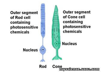
 · Retina layer It is the innermost layer of the eye which is similar to the film of the camera.It changes the light into images and transfers to the brain via optic nerve. The two types of photoreceptors in this are § Blind spot: Rods and Cones are absent hence light cannot carry to the brain. § Yellow spot (Fovea centralis): Rods are absent and Cones are present hence only clear vision is carried to the brain. Retina contains two major types of light sensitive photoreceptor cells used for vision Differences between rods & cones Rods | Cones | They cannot differentiate the colors but are responsible for the scoptic and monochromic vision. | They can differentiate the colors and are responsible for the coloured vision. | They work well in dim light because of rhodopsin pigment. | They work well in bright light because of a pigment iodopsin. | They are present in the retina but moves at the fovea and blind spot. | They are present in and near to the fovea and few are in the side of the retina. | Example: Owls | Example: Sparrows |
Ø Chambers of the Eye ball The lens, ciliary body and suspensory ligament divides the eye ball into two chambers namely: Differences between aqueous & vitreous chambers Aqueous Chamber | Vitreous Chamber | It is a transparent, gelatinous b fluid secreted from the ciliary body supports the lens. | It is a transparent, gelatinous mass secreted in the retinal cells supports the lens and retina. | It is located in between the lens and cornea of the eye. | It is located in between the lens and retina of the eye. | Function: supplying nutrition to the lens and cornea. | Function: maintaining the shape of the eye ball. |
Ø Lachrymal glands These glands which are associated with water water which are present in the eyes by secreting watery salt liquid helps in destroying bacteria, keeping the eye moist and removing the dust particles. In some emotional states the fluid flows externally and spills over known as tears hence also known as tear glands. Ø Defects of the Vision · Short Sightedness The person with this defect was unable to see the distant objects clearly because the light is focused in front the retina. o Treatment: Using spectacles with concave diverging lenses. · Long Sightedness The person with this defect was unavle to see the near objects clearlky because the light is focused behind the retina, o Treatment: Using spectacles with concave converging lenses. · Cataract The lens of the person becomes cloudy or opaque if any injury occurs. They will prevent the light rays reaching to the retina and finally leads to blindness. o Treatment: Using special eye glasses and removing the cloudy lens by surgery. · Night Blindness(Xerophthalmia) The person was unable to see the object clearly in the dim light because the synthesis of rhodopsin pigment decreases. o Treatment: Eating leafy vegetables. · Colour blindness It is a disease due to the absence of cones in the retina. o Treatment: Nil.
|